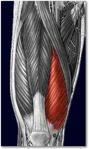
Anatomy
| Parts | Rectus femoris, vastus lateralis, vastus medialis, vastus intermedius |
| Origins | Rectus femoris: Anterior inferior iliac spine, supraacetabular groove Vastus medialis: Intertrochanteric line, pectineal line of femur, linea aspera, medial supracondylar line of femur Vastus lateralis: Intertrochanteric line, greater trochanter, gluteal tuberosity, linea aspera of femur Vastus intermedius: Anterior surface of femoral shaft |
| Insertions | Rectus femoris and vastus intermedius: Tibial tuberosity (via patellar ligament), patella Vastus lateralis: Tibial tuberosity (via patellar ligament), patella, (lateral condyle of tibia) Vastus medialis: Tibial tuberosity (via patellar ligament), patella, (medial condyle of tibia) |
| Innervation | Femoral nerve (L2-L4) |
| Function | Hip joint: Thigh flexion (rectus femoris only); Knee joint: Leg extension |
Anatomy
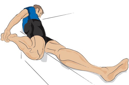
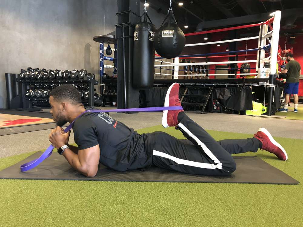
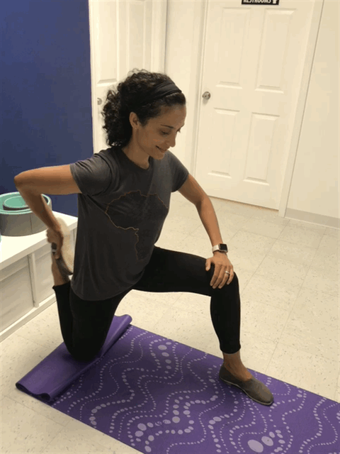
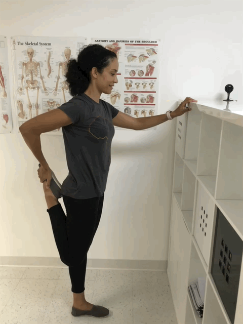
Stretching
Q (Quadriceps ) Angle
Note: The Q angle is the angle between the quadriceps tendon and the patellar tendon. The Q-angle is formed from a line drawn from the ASIS to the center of the kneecap, and from the center of the kneecap to the tibial tubercle. To find the Q-angle, measure that angle, and subtract from 180 degrees.
To Measure Q Angle:
- Patient is standing, with the knee in extension and
- femur neutral: (no internal or external rotation) and
- patient’s feet in a neutral position (no pronation or supination)
Normal Q Angle Test Result:
A normal Q angle with the knee extended and the quadriceps muscle relaxed is 18° degrees for women and 13° degrees for men.
A Q angle that is less than normal allows the patella to track medially between femoral condyles, placing extra stress on the medial articulating facets of the patella which leads to Chondromalacia Patellae
A Q angle that is greater than normal allows the patella to track laterally, stressing the lateral facets which is associated with patellar tracking dysfunction, chondromalacia patellae and patellar subluxation.
quadriceps
